
Salt Lake City – The National Science Foundation (NSF) and the journal Science have awarded top honors to Bryan William Jones, Ph.D. of the John Moran Eye Center in the 2012 International Science & Engineering Visualization Challenge. Jones received first place in the photography category for his image of a metabolic snapshot of a mammalian retina which will be featured in the upcoming issue of Science and Science Online available on February 3, 2012.
The International Science & Engineering Visualization Challenge was created to celebrate illustrations that provide the most immediate and influential connection between scientists and other citizens, with the hope for nurturing popular interest. They are a necessity for public understanding of research developments. The spirit of the competition is for communicating science, engineering and technology for education and journalistic purposes.
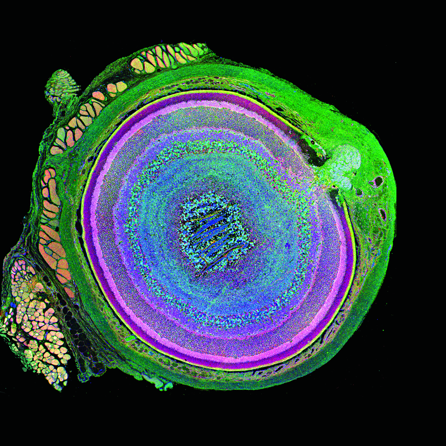
Jones’ image was made possible because of a technique called computational molecular phenotyping or (CMP) that was originally developed by Dr. Robert E. Marc, the director of research at the Moran Eye Center. CMP reveals complex metabolic signals in all cells of tissues while preserving the anatomical context and providing insight into the metabolic diversity of eyes, which are composed of over 70 types of cells.
Using a special kind of precision knife called an ultramicrotome, Drs. Jones and Marc machined then serially sectioned a mouse eye at the nanometer scale to probe it with antibodies against specific small molecules of interest. In this case, three separate 200nm thick sections were probed with taurine, glutamine and glutamate and the resulting data was assigned to red, green and blue color channels respectively. Some of the muscles that move the eye can be seen on the outer, leftmost portion of the image as a golden color while the sclera which is normally the white part of your eye is shown in green (see image). Other classes of cells that make up the eye including epithelia, neurons and glia are represented in different combinations of reds, blues, pinks, yellows and orange.
“Scientifically, this image not only shows the diversity of cell types in complex tissues like the retina, but also demonstrates that cells within a class type possess identical metabolic ‘fingerprints’ that can be used to track cell identity and potentially serve as a new tool to identify and track disease processes in everything from retinal degeneration to diabetes and cancer,” says Jones.
About Bryan Jones, Ph.D.
Bryan William Jones joined the Research Faculty of the Moran Eye Center in 2006. Originally coming to science through the study of epilepsy and sleep medicine, he became fascinated by the beauty of the retina and the complexity of blinding diseases. His work with Dr. Robert E. Marc pioneered our understanding of retinal remodeling by revealing the nature and extent of pathology seen in retinal degenerative diseases such as retinitis pigmentosa and macular degeneration. This work has helped to refine approaches to vision rescue through both bionic and biological approaches. Continued work in retinal degenerative diseases will further define the time lines of retinal remodeling in an effort to determine windows of opportunity for intervention to limit, prevent or exploit retinal remodeling. Dr. Jones other work with Dr. Marc focuses on applying novel molecular and computational approaches to rescuing vision loss, resolving the identities and connectivities of retinal neurons to discover how the normal retina is wired and how that circuitry is altered in degenerative diseases.
About the John A. Moran Eye Center
The John A. Moran Eye Center is committed to the goal that no person with a blinding condition, eye disease or visual impairment should be without hope, understanding, and treatment. The Moran Eye Center is home to more than 50 faculty members, including one of the top retinal research teams in the world. Moran researchers are largely funded by grants from the National Institutes of Health and are involved in more than 50 active clinical trials. Each year, Moran hosts more than 120,000 clinic visits, including more than 7,000 surgeries. Ophthalmologists at the Moran Eye Center are highly involved in international outreach and perform multiple medical missions throughout the world every year.